|
Abstract: A 67-year old diabetic patient showed yellowish discoloration and dystrophy of several fingernails. Mycological examination revealed infection with Trichophyton gallinae. This is the first report of human onychomycosis caused by this zoophilic fungus in Germany. Key words: fungal infection, nails, onychomycosis, zoophilic dermatophytes, mycoses, Trichophyton gallinae, Microsporum gallinae (Syn.), Achorion gallinae (Syn.). |
A 67-year old female patient complained of itching rash of forearms, which had persisted for 2 years. She had diabetes, for which she was taking insulin and glibenclamide. There were no other medications, neither history of previous allergic reactions.
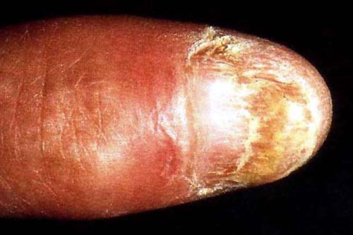
There were single excoriated papules and pigmented macules on the ventral sides of both forearms. The examination also revealed yellow-grey discoloration of the fingernails and onycholysis on the left hand (Fig. 21). Toenails were not involved.
Fig. 21: Onychodystrophy of the left hand's thumbnail.
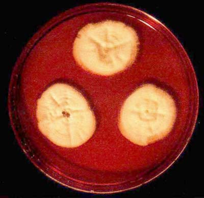
KOH examination of the diseased fingernail fragments was negative. Samples of the nail were cut into small pieces and inoculated on Sabouraud agar at 24°C for 3 weeks. Based on the appearance of the characteristic reddish colour in the culture plate, as well as macroscopic and microscopic features, the zoophilic fungus Trichophyton gallinae was identifies (Fig. 22).
Fig. 22: Colonies of Trichophyton gallinae.
Repeated questioning of the patient revealed a possible source of the zoophilic infection: The patient had contact with carrier pigeons bred by her husband. Systemic antimycotic therapy was planned, however, it could not be undertaken due to hepatic steatosis with persistent elevation of transaminases. The skin changes on the forearms were diagnosed as a diabetes-related pruritic dermatosis, and treated respectively. The patient did not appear for further antimycotic treatment.
Dermatophytes are pathogenic fungi capable of digesting animal or human keratin and using it as nutrient source. The dermatophyte Trichophyton gallinae is a bird pathogen, which very seldom infects humans or other mammals. This is due to specificity of the enzyme keratinase of the fungus - it can digest keratin of bird feathers but hardly that of the mammalian hair (1). Only a few exceptions from this rule are known: In individual cases, T. gallinae was isolated from apes and dogs (2, 3). Faruqui et al. reported on a Pakistani child with tinea capitis caused by T. gallinae (4). In Iran, the frequency of mycoses caused by T. gallinae among humans was estimated at 0.2% (5). Interestingly, the fungus seems widespread among people in Nigeria, where recently 18.4% of all mycoses among children were attributed to T. gallinae (6).
Only a few cases of human T. gallinae-infection were reported from Europe. In 1964, two men with tinea cruris were described in Sweden, there was also a case of occupational infection in a Bulgarian farmer (7, 8). In 1984, Arievich et al. reported on a teacher who contracted the infection from chicks kept as pets in one of Moscow's kindergartens (9). Herpetiform tinea due to this fungus was described in a French child, also in this case the infection was transmitted from a pet hen kept at home (10). In all above-mentioned cases, the mycosis had a localised form, only one case of generalised T. gallinae-infection was reported in a Spanish AIDS patient (11).
It appears that human T. gallinae-infections are typically contracted from livestock birds, especially chickens and pigeons. In Central Europe, however, zoophilic dermatophytoses are generally extremely rare, and not a single case of T. gallinae-infection was observed so far among farmers or animal breeders (12, 13). To our best knowledge, this is the first case of T. gallinae-onychomycosis in Germany.
For personal use only. © Blackwell Wissenschafts-Verlag GmbH.
| Contact Dr. Spiewak | Back to article list | Website's front page |